The laryngoscope line-up
Spin through six scopes, from 1895 to today. Tap a scope, or use the button under it, to jump to its story below.
The curve follows the tongue, and the tip lifts the epiglottis from the vallecula.
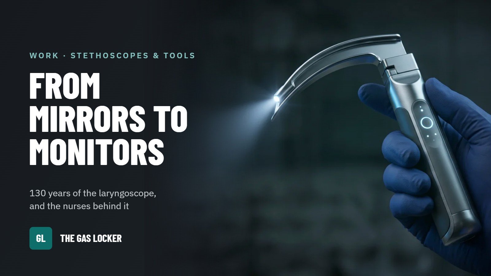
Every CRNA reaches for a laryngoscope without thinking about it. That handle and blade carry 130 years of trial and error, and nurse anesthetists were part of the story almost from the start. Here is a short look at the nurses who built the profession, then a scope-by-scope walk from the first direct look at the larynx to the screens we use today. Tap any scope in the line-up to jump to its history.
Key takeaways
- Nurses have given anesthesia in America for more than 150 years, starting with Sister Mary Bernard in 1877.
- Agatha Hodgins founded today’s AANA in 1931, the first national association of anesthesia providers in the U.S.
- Kirstein took the first direct look at the vocal cords in 1895; the Miller (1941) and Macintosh (1943) blades are still used today.
- The GlideScope became the first commercial video laryngoscope in 2001.
Last reviewed Oct 2026 · Written by a practicing CRNA
A brief history of nurse anesthesia
Nurses have been giving anesthesia in America for more than 150 years, and they organized nationally before any other anesthesia providers did.
We’ve done it before, and we can do it again.
Founding Mothers
Sister Mary Bernard
In 1877, Sister Mary Bernard became the first nurse to specialize in anesthesia, at St. Vincent’s Hospital in Erie, Pennsylvania (AANA).
Alice Magaw, the “Mother of Anesthesia”
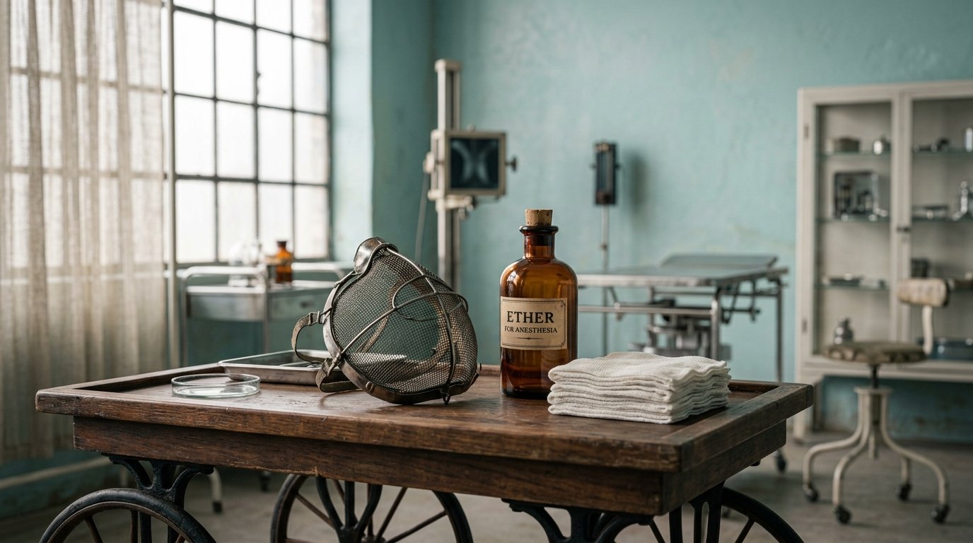
Alice Magaw gave anesthesia at St. Mary’s Hospital in Rochester, Minnesota, working with Dr. Charles Mayo in the 1890s, in the years that grew into the Mayo Clinic (AANA). Her skill with open-drop ether led Dr. Mayo to call her the “Mother of Anesthesia.” She recorded more than 14,000 anesthetics without a single anesthesia death (Working Nurse). She also published her results, which helped other hospitals copy her methods.
Agatha Hodgins
Agatha Hodgins, a Canadian-born nurse, taught anesthesia to nurses and doctors at Lakeside Hospital in Cleveland, Ohio, from 1911. In 1915 she formally opened the Lakeside Hospital School of Anesthesia (AANA).
On June 17, 1931, she founded the National Association of Nurse Anesthetists in Cleveland. It was the first national association of anesthesia providers in the U.S., and it is today’s AANA (AANA).
Nurses at War
In the noise of war, the calmest presence in the room was often a nurse, caring for each soldier the way a mother would.
The Civil War
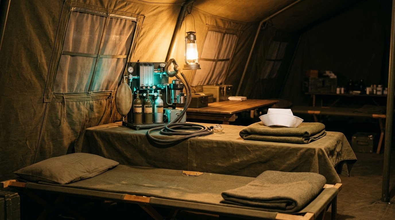
Nurse anesthesia started on the battlefield. During the American Civil War, nurses began giving anesthesia to wounded soldiers. Some military surgeons noticed that nurses watched the whole patient, while medical students were more interested in the surgery itself (Working Nurse).
World War II
World War II put nurse anesthetists on every front. The Army Nurse Corps grew from fewer than 1,000 nurses in 1941 to about 59,000 by the end of the war, and more than 2,000 of them were trained to give anesthesia (AANA Journal).
They worked in evacuation hospitals across North Africa, Italy and the Pacific, often with ether, thiopental and nitrous oxide. Adeline Simonson of the 95th Evacuation Hospital, called “our super competent anesthetist” by a colleague, moved with her hospital from Morocco to Salerno and Anzio. At Anzio, a German bombing on February 7, 1944 killed 22 of the hospital’s staff, including three nurses. In Hawaii, nurse anesthetist Mildred Irene Clark trained anesthetists for deployment. She later became the first nurse anesthetist to lead the Army Nurse Corps, in 1963 (AANA).
The evolution of the scope
Before 1895, the only way to see the vocal cords was with mirrors. In 1854, Manuel García used two mirrors and sunlight to see a living larynx at work (Wikipedia). Everything below is the story of looking straight in instead.
1895: Kirstein’s autoscope
On April 23, 1895, Alfred Kirstein of Berlin described the first direct look at the vocal cords (Wikipedia). He used a modified esophagoscope he called an autoscope, with the light at the handle end.
The discovery was partly an accident. A colleague had slipped an esophagoscope into a patient’s trachea and got “a perfect view of the bronchial bifurcation” (EMS World). Kirstein realized he could look straight down at the larynx.
1913: Jackson’s lighted blade
Chevalier Jackson, professor of laryngology at Jefferson Medical College in Philadelphia, moved the light to the tip of the blade in 1913 (ENT Today). His blade had a sliding floor that made room for an endotracheal tube or a bronchoscope.
Jackson was the first to report a high success rate for intubating through direct laryngoscopy (Wikipedia). He was also famous for removing more than 2,000 swallowed objects from patients’ airways (EMS World). The same year, Henry Janeway added batteries in the handle, a step toward the scopes we use now.
1941: The Miller blade
Anesthesiologist Robert Arden Miller introduced his blade in 1941, as intubation was becoming routine (Wood Library-Museum). It is a straight blade with a long, curved tip, a small light built into the blade, and a detachable blade on a battery handle.
The design made hard intubations easier in the years before muscle relaxants were common. It first came only in a medium size. Today it runs from size 0, for premature infants, up to size 5. It remains the most popular straight blade (Wikipedia).
1943: The Macintosh blade
Sir Robert Macintosh, Britain’s first professor of anesthesia at Oxford, introduced his curved blade in 1943 with technician Richard Salt (Wood Library-Museum). Instead of lifting the epiglottis directly, the tip sits in the vallecula. Lifting the handle raises the base of the tongue, and the epiglottis comes up with it. A flange on the left edge sweeps the tongue aside.
The idea came by accident during a tonsillectomy list. A Boyle-Davis mouth gag left the cords “perfectly displayed,” so Salt soldered a gag blade onto a laryngoscope handle (LITFL).
Macintosh never patented it or took royalties, so manufacturers everywhere copied it. The hook-on blade attachment followed, and by 1952 the manufacturer Foregger had switched to it entirely (LITFL). One historian called it “probably the most successful ‘durable’ in the history of anaesthesia.” It is still one of the most popular blades in the world.
1967: The flexible fiberscope
In 1967, Peter Murphy performed the first fiber-optic nasal intubation, using a fiber-optic choledochoscope in a patient with Still’s disease (PMC review). The scope could bend around the curve of the airway, and the tube rode over it into the trachea.
By 1972, Stiles and colleagues had reported fiber-optic intubation in 100 patients, and the technique spread to difficult airways in adults and children.
2001: The video laryngoscope
In 2001, the GlideScope became the first commercially available video laryngoscope (Wikipedia). It was designed by Dr. John Pacey, a vascular and general surgeon, after a prototype in 1999. A camera near the tip sends the view to a screen, so the provider sees around the curve instead of needing a straight line of sight.
Video laryngoscopes now come with standard Mac-style blades and steeper hyperangulated blades. For how they fit into a surgery center today, read our video laryngoscope guide.
What hasn’t changed
The tools went from mirrors to monitors, but the job at the head of the bed stayed the same: watch the whole patient and secure the airway. Alice Magaw did it with open-drop ether. WWII nurse anesthetists did it in evacuation hospitals near the front. CRNAs do it today with a screen in one hand. The scope keeps changing. The person holding it is the constant.
For today’s devices in practice, see Video Laryngoscopes in the ASC.
Sources
- AANA: History of Nurse Anesthesia
- Working Nurse: 130 Years of Anesthesia Nursing
- AANA Journal, October 2016: Imagining in Time (WWII nurse anesthetists)
- Wikipedia: Laryngoscopy
- ENT Today: History of the Laryngoscope
- EMS World: Great Intubators and Innovators
- Wood Library-Museum: Miller Laryngoscope
- Wood Library-Museum: Macintosh Laryngoscope (prototype)
- LITFL: Macintosh Laryngoscope
- PMC: Teaching and training in fibreoptic bronchoscope-guided endotracheal intubation
Educational history, not clinical instruction. The 3D scopes are generic illustrations, not specific brands or products. Cover image: AI-generated via Google Gemini. This is educational history, not clinical instruction. There are no affiliate links in this article. Image credits: Image: AI-generated via Google Gemini.